
Diagnostic Cardiology
What is the connection between sleep medicine and cardiology?
Unrefreshing sleep can not only cause subjective effects, such as daytime sleepiness and poor concentration, but also affects the cardiovascular system.
The sleep apnea syndrome which is characterized by repeated breathing arrests carries a high risk for cardiovascular disease. If breathing is interrupted during sleep, there is a drop in oxygen saturation in the blood, and hard breathing triggers a stress and wake-up reaction. The circulatory system will be slowed down and the blood pressure rises. Repeated severe high blood pressure, in turn strains the heart muscle. The regular nightly strain may cause heart failure and cardiac arrhythmia.
A sleep apnea, even milder forms, is to be sought mainly in the presence of the following disorders:
- Circulatory / stroke disorders of the arteries (neck, heart, legs ...)
- Heart attack, stroke
- Intermittent claudication
- Cardiac arrhythmia of any kind
- Aortenenuerysma (aneurysm of the aorta in the chest or the abdomen)
- High blood pressure (hypertension)
- Diabetes and impaired glucose metabolism
Treating sleep apnea leads to a decrease in the disease or disorder or its progression. When other factors are implicated in the pathogenesis of the disease, the disorder can be treated and monitored more easily.
For the diagnosis of cardiac disorders the following methods and equipment are available in our practice:
Resting ECG
With the help of electrodes, the heart's electrical impulses are recorded, resulting from any action of the heart.
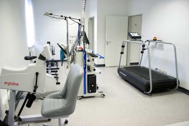
Exercise ECG
Registration of the ECG and blood pressure under gradually increasing load on the seat or semi-prone ergometer. This serves the diagnostics load-dependent disorders.
Holter ECG
Registration of the ECG with a digital mini-recorder for 24 hours, if necessary with cardiac pacing detection, analysis and detection of cardiac arrhythmias.
Blood pressure measurement, and SphygmoCor (proximal cardiac blood pressure)
The measurement of blood pressure is usually performed with a cuff and a stethoscope to the brachial artery or wrist. However, these measured values in some cases significantly deviate from the central aortic pressure. More precise measurements can be obtained with the new SphygmoCor system. It provides a simple and noninvasive pulse wave analysis of the aortic pressure near the heart. A pen-like sensor can be configured via a computer program which calculates and represents heart pulse waves. Thus, early forms in blood pressure and vascular changes can be detected early.
Cardiac ultrasound (echocardiography)
The examination with modern, high-performance devices enables the assessment of heart size, wall thickness and size of the chambers of the heart valves as well as function and structure of large arteries. The direction of blood flow of can be shown by Doppler echocardiography.
Stress echocardiography
Ultrasound assesses the cardiac function by gradually increasing load. This is part of an exercise ECG done on a semi-prone ergometer, which may be tilted in a left lateral position.
Ultrasound of the vessels
The arteries in the neck are close to the surface. This allows a good representation of the blood flow and clogging of the vessels and the structure of the vessel wall. Changes in the walls, which arise as a consequence of cholesterol and fat deposits can already be detected both in early or advanced stage of calcification. Thus, the extent of atherosclerosis can be assessed. The method therefore serves the risk evaluation and assessment of the likelihood of a stroke.
Computed tomography of the heart:
Using computed tomography, the heart can be displayed three-dimensionally in high resolution. Even the small coronary arteries can be evaluated very carefully. Early changes and deposits can be detected. If calcifications are already there, the process of vascular calcification in the coronary arteries is more advanced and circulatory problems are already possible. Due to the high sensitivity of the method there is an early detection of cardiovascular calcifications (coronary heart disease) and risk assessment.
Magnetic resonance imaging of the heart (cardiac MRI)
With the MRI, the heart can be imaged without X-rays. In addition, the structure and properties of the heart tissue can be assessed. Detailed information about the presence of inflammation, scar tissue or circulation problems can be given. The advantage of the MRI is that it is not based on X-rays. It is harmless magnetic fields and radio frequency waves.